Hemostatics: Materials and Applications
An Introduction to Hemostasis
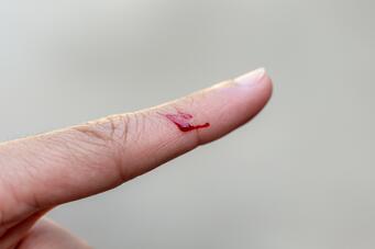
Accidents happen quickly—one moment of carelessness, and we might cut ourselves, perhaps while cooking. Though painful and often accompanied by bleeding, depending on the depth of the cut, we typically notice the blood flow slowing and eventually stopping. By the next day, the wound is usually closed, and within a few more days, only a faint red mark remains. This natural process, begins with blood coagulation, known as hemostasis, which works to stop the bleeding. While this process is sufficient for many minor injuries, excessive blood loss remains the leading cause of trauma-related deaths.[1]

Basic Hemostatic Methods: Gauze and Pressure
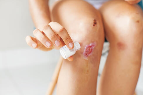
To prevent severe blood loss in both clinical settings and first-aid situations, various hemostatic measures are employed. The most basic method for supporting the body's natural hemostatic response involves wound closure and applying pressure. Gauzes, made from different materials, are effective for many types of wounds.
However, when these measures are either insufficient or inapplicable, other hemostatic agents are needed.
Advanced Hemostatic Agents: When Basic Methods Fall Short
For example, in surgeries where traditional gauze and pressure are not viable options—such as when operating on internal organs—materials are required that can safely remain inside the body post-operation. One approach supports the natural hemostatic pathway by introducing fibrinogen and thrombin. These are both key components of the hemostatic cascade, in which fibrinogen is converted into fibrin through the action of thrombin and other coagulation factors. However, because these substances are often derived from human blood plasma, there is a risk of transmitting infections from donors.[2]
In oral surgery, aluminum chloride solution is frequently used due to its acidic, astringent, and protein-binding properties, which help promote blood coagulation and accelerate hemostasis.[3]
Another method involves sealing wounds with cyanoacrylate adhesives. While these adhesives provide a fast solution, they come with drawbacks such as heat generation during curing and the release of potentially toxic degradation products.[4]


Biopolymers: Hemostatics for Internal and Topical Application
Less chemically invasive approaches include hemostatic powders made from absorbent biopolymers, such as starch or modified starch. These particles, with their high absorption capacity, clear the surgical site of excess blood. Acting like molecular sieves, they promote blood coagulation by absorbing blood plasma and thereby increasing the local concentration of platelets. While other hemostatic powder such as powders from oxidized cellulose exist, starch-based materials have the advantage of better bioresorption and, depending on the specific formulation, can be naturally cleared from the surgical site within a day.[5]
In addition to their use in surgical settings, fine powders are valuable for treating topical wounds, especially those with large surface areas or unclear bleeding sources.
In topical applications powders and gauzes made from chitosan have gained significant attention in recent years for their hemostatic properties. Chitosan, a biopolymer derived from crustacean or fungal chitin, combines excellent biocompatibility with versatility, as it can be processed into fine fibers, powders, or gels. Its advantage over basic gauze lies in its added hemostatic properties: chitosan promotes platelet aggregation and plasma protein coagulation. After aiding clot formation, it further enhances wound healing by stimulating the activity of various cellular processes.[4] Additionally, chitosan is of particular interest because of its synergistic effects when combined with other wound-healing and hemostatic biomaterials, such as calcium alginate.[6]
Conclusion
While the human body has an intricate and effective hemostatic system, severe trauma and medical conditions can overwhelm it, requiring external support. Biomaterials have emerged as key players in enhancing and accelerating the body’s natural hemostatic process, offering solutions where traditional methods like gauze and pressure fall short.
Modern biomaterials provide a versatile toolkit for managing bleeding in both clinical and emergency settings. Their ability to mimic or enhance natural hemostasis, while being biocompatible and bioresorbable, positions them as vital components in the future of wound care and surgery. As research continues, the potential to develop even more sophisticated biomaterial-based solutions remains vast, promising to address the challenges of complex bleeding scenarios with greater efficiency and safety.
In highlighting these advances, we see biomaterials not only as a supplement to traditional hemostatic methods but as a transformative force that can redefine how we approach wound management and surgical care.
Join the Conversation
What are your thoughts? Where do you see the limitations and possibilities of modern biomaterials in hemostasis?
We invite you to connect with us—let’s explore the future of hemostatics together.
[1] Teixeira et al.: Preventable or Potentially Preventable Mortality at a Mature Trauma Center. The Journal of Trauma: Injury, Infection, and Critical Care 63(6):p 1338-1347, December 2007. DOI: 10.1097/TA.0b013e31815078ae
[2] Daud et al.: Fibrin and Thrombin Sealants in Vascular and Cardiac Surgery: A Systematic Review and Meta-analysis, European Journal of Vascular and Endovascular Surgery,Volume 60, Issue 3,2020,Pages 469-478,ISSN 1078-5884,DOI: 10.1016/j.ejvs.2020.05.016
[3] Tarighi et al.: A review on common chemical hemostatic agents in restorative dentistry. Dent Res J (Isfahan). 2014 Jul;11(4):423-8. PMID: 25225553; PMCID: PMC4163818.
[4] Fan et al.: Chitosan-Based Hemostatic Hydrogels: The Concept, Mechanism, Application, and Prospects. Molecules. 2023 Feb 3;28(3):1473. doi: 10.3390/molecules28031473. PMID: 36771141; PMCID: PMC9921727.
[5] LyBarger (2024) Review of Evidence Supporting the Arista™ Absorbable Powder Hemostat, Medical Devices: Evidence and Research, 17:, 173188, DOI: 10.2147/MDER.S442944
[6] Xiaowei et al.: Fabrication of chitosan calcium alginate microspheres with porous core and compact shell, and application as a quick traumatic hemostat, Carbohydrate Polymers, Volume 247, 2020, 116669, ISSN 0144-8617, https://doi.org/10.1016/j.carbpol.2020.116669.


